

Complex brain tumor removal gives medical student a second chance

Medical student Cherechi Abakwue, 29, is on track to graduate from Loma Linda University in California thanks to care she received from the UTHealth Houston Skull Base Program. (Photo by Cherechi Abakwue)
In 2023, Cherechi Abakwue, now 29, was only a few months into her first year of medical school at Loma Linda University in California when she began experiencing headaches.
The headaches, which began just before she turned 27, came on when she laughed or strained her voice, but she chalked it up to the stress of the intense study sessions required by medical school. But by the next summer, the headaches were no longer something she could ignore. She scheduled an appointment with her California doctor, who ordered an MRI shortly after the July 4 holiday.
Four hours after the appointment, Cherechi received a call that made her world stand still. The MRI revealed she had a tumor the size of a small orange—or about three to four fingers wide—that was pressing on her brainstem.
“I just remember in the middle of my call, I looked down at my fingers and was like, ‘Oh, no. This might be it for me,’” Cherechi said.
Cherechi’s tumor was a meningioma, the most common type of primary brain tumor that develops on the tissue surrounding the brain and spinal cord. Meningiomas are more common in women, in part because of the hormonal receptors for estrogen and progesterone.
While benign, Cherechi’s meningioma was pressing on her brainstem—a dense and critical nerve tract that controls everything in a person’s neural wiring, including the ability to move, speak, walk, eat, and breathe.
Even with the knowledge she’d gained in medical school, she was unprepared to be handed such a severe diagnosis.
“Whenever you’re in a medical situation, even as a medical student, you blank out, and you just don't remember anything,” Cherechi said. “I just couldn’t remember, is this good? Is this bad? Is this cancer?”
The reality didn’t fully set in until Cherechi followed up with a California neurosurgeon, who showed her images from her MRI.
“I honestly didn’t think it was me until I started looking at the person in the MRI and was like, ‘That's my nose. That’s my lips. That’s my big old head. This is actually me,’” Cherechi said.
When Cherechi told her doctor that she was a Houston native, she was referred to Jacques J. Morcos, MD, an expert in skull base tumors and cerebrovascular surgery who is a professor and John P. and Kathrine G. McGovern Distinguished Chair in the Vivian L. Smith Department of Neurosurgery at McGovern Medical School at UTHealth Houston.
“Her doctor called me from California to tell me, ‘I’m sending you this patient. This requires your expertise,’” said Morcos, who is also co-director of UTHealth Houston Neurosciences.
Morcos confirmed Cherechi’s diagnosis and determined she required treatment through the UTHealth Houston Skull Base Program, a multidisciplinary team that focuses on complex brain tumors and other conditions. The program was recently designated a North American Skull Base Society Multidisciplinary Team of Distinction for 2026.
“The skull base is a border zone between the brain, the face, the neck, the ears, and the eyes. By definition, it’s more complex because there’s lots of stuff there: brainstem, nerves, arteries, veins, muscles,” Morcos said. “Cherechi’s tumor is actually the hardest, most complex tumor to have. It is known as a petroclival meningioma.”
Things started to click for Cherechi after speaking with Morcos. She noticed other symptoms, like having trouble swallowing and maintaining her balance. She thought back to months before, when she had unexpectedly tripped while looking through her closet, an incident that was confusing at the time but now had an explanation.

Cherechi Abakwue, 29, at the Student National Medical Association's Annual Medical Education Conference. (Photo by Cherechi Abakwue)
Her surgery was scheduled for Aug. 22, 2024. Because of the location of the tumor, a host of complications were possible during surgery, including quadriplegia, blindness, loss of hearing, or a coma. Cherechi was terrified.
“I thought, if I were to die, I’m not going to know that I’m dead. But my family will be so distraught. I just don’t want them to be so sad without the funniest girl in the family, you know? What am I gonna do? Who’s gonna make them laugh?” she said. “I vividly remember whenever they rolled me back, I was like, ‘This feels like a funeral, except that I’m awake.’”
To complete the procedure, Morcos required the expertise of Vivian F. Kaul, MD, assistant professor and chief of Otology & Neurotology at UTHealth Houston. Kaul made a C-shaped incision behind Cherechi’s right ear and drilled into the skull bone, allowing Morcos to access and remove Cherechi’s tumor.
Throughout the surgery, Morcos conducted neurophysiological monitoring of Cherechi’s brain and nerve functions to ensure she was not being harmed while the tumor was being removed.
“A technician in the room placed electrodes and wires in specific areas of her face, her head, her body,” Morcos said. “He continuously stimulates various brain and nerve functions and makes sure the tracing doesn’t weaken. As long as the tracing remains at her baseline, it means those nerves, those functions are working, and that I, as a surgeon, am not hurting anything.”
Ten hours later, Cherechi was awake in her post-op room.
“I remember waking up and thinking ‘I’m alive,’ even though I didn’t know anything else. I was so grateful to God, and I was so grateful to Dr. Morcos, the whole team,” she said.
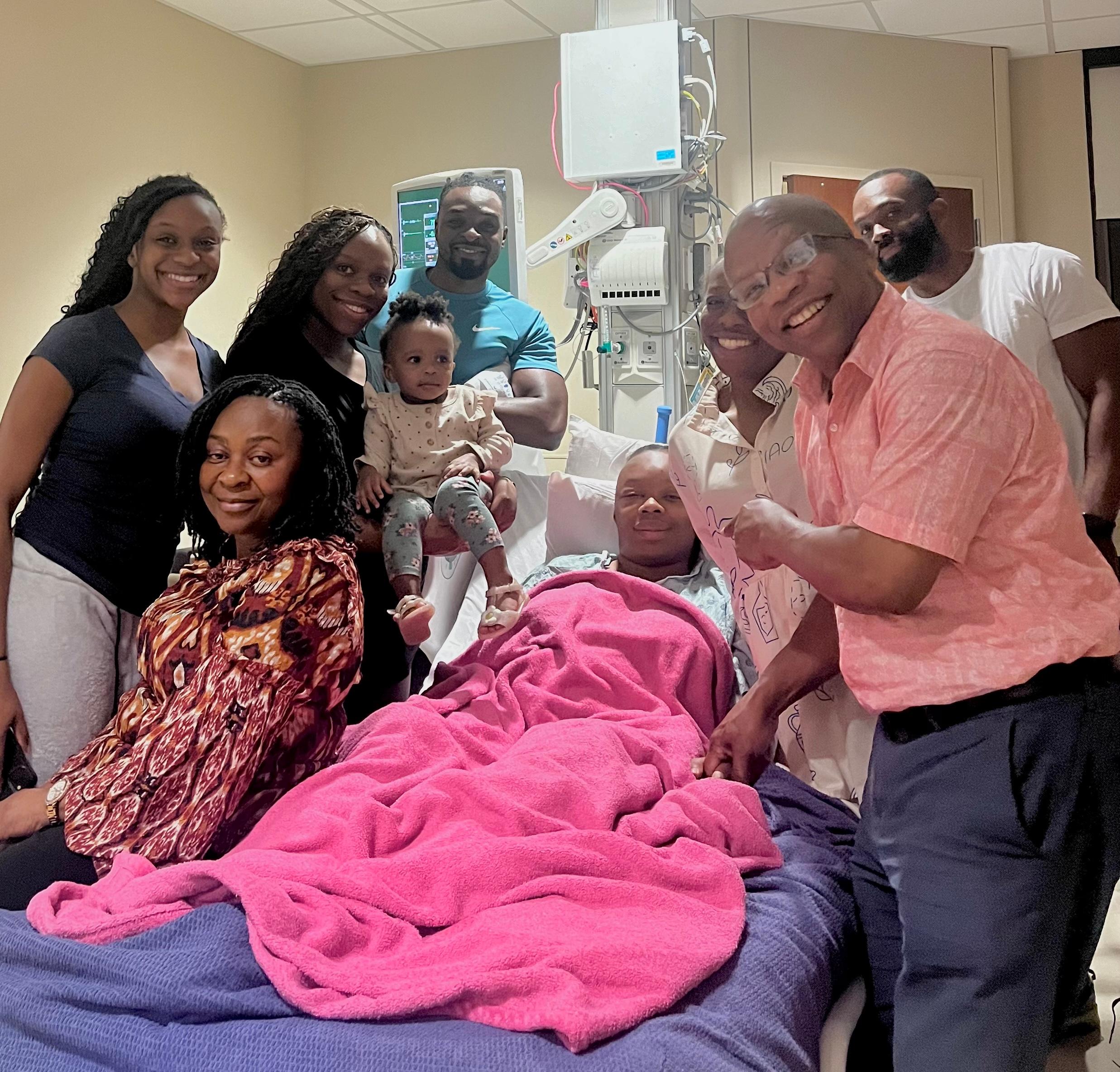
Cherechi Abakwue, 29, surrounded by family as she recovers from a 10-hour brain surgery performed by Jacques J. Morcos, MD. (Photo by Cherechi Abakwue)
Cherechi’s surgery went remarkably well, with only two minor complications. Because of the location of her tumor, Morcos had to sacrifice her fourth cranial nerve, which connects to one of the muscles in the eye. Cherechi will have to live with slight double vision out of her right eye, which she can compensate for and that can be fixed with prism glasses or minor corrective eye surgery.
A few weeks after the operation, Cherechi developed a pseudomeningocele, which occurs when cerebrospinal fluid leaks from the brain and spinal cord into surrounding tissues. Morcos placed a small, catheter-like tube known as a lumbar drain in Cherechi’s lower spine to drain the fluid. After several days with the drain, the pseudomeningocele resolved without the need for another brain surgery.
By December 2024, Cherechi was back at Loma Linda University and caught up on her second year of medical school, an accomplishment she credits to Morcos.
“Dr. Morcos reached out to the dean of my school during my first appointment. It was phenomenal,” Cherechi said. “There was something about Dr. Morcos that felt God-sent. He made me feel like I was his only patient.”
As she enters her final year of medical school, Cherechi says her journey with Morcos has inspired her own approach to medicine.
“With me and Dr. Morcos’ relationship, yes, I had a brain tumor. Yes, I knew I had to get it out. But my issue was I didn’t want to miss any more school,” Cherechi said. “Dr. Morcos said, ‘Give me the names.’ He reached out to the dean of my school, anybody I wanted him to reach out to. That was healthcare for me. It made me look beyond and think, if I was a patient in this position, what would I want? That’s how I plan on practicing medicine, with compassion.”


